Diagram Neck Anatomy Glands - Neck Anatomy Robins Levels Neck Dissection Posterior Triangle Anatomy Tutorial Youtube - Some important structures contained in or passing through the neck include the seven cervical vertebrae and enclosed spinal cord, the jugular veins and carotid arteries, part of the esophagus, the larynx.
byAdmin-
0
Diagram Neck Anatomy Glands - Neck Anatomy Robins Levels Neck Dissection Posterior Triangle Anatomy Tutorial Youtube - Some important structures contained in or passing through the neck include the seven cervical vertebrae and enclosed spinal cord, the jugular veins and carotid arteries, part of the esophagus, the larynx.. The submaxillary glands (lymphoglandulæ submaxillares) (fig. Specific sub sites of these organs, which are considered lateral sites, are indicated with an asterisk (*) in the code table above. These include hair, sweat glands, sebaceous glands, and sensory nerves. The anatomy of the head and neck of the human body, including the bones, muscles, blood vessels, nerves, glands, nose, mouth, and throat shoulder anatomy diagram. Extending from underneath the chin, to the posterior aspect of the head.
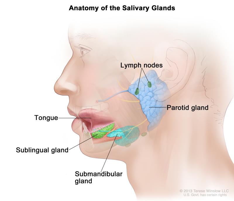
Apart from the main glands, there are also a number of other salivary glands whose main purpose is to keep the inner lining of the oral cavity moist and lubricated at all times. The throat anatomy tonsils are located over clefts of tissues in the rear part of the throat. The submaxillary glands (lymphoglandulæ submaxillares) (fig. Ear nose and throat medical illustrations. 604), three to six in number, are placed beneath the body of the mandible in the submaxillary triangle, and rest on the superficial surface of the submaxillary salivary gland.
The Salivory Glands Medika Life Understanding Human Anatomy from medika.life They drain your conjunctiva, lips, tongue, and flour of your mouth. The tonsils also are lymphatic tissue and help mediate the ingestion of pathogens. Related posts of diagram of the neck anatomy veins and arteries of the neck. The neck is the part of the body, on many vertebrates, that separates the head from the torso. Some important structures contained in or passing through the neck include the seven cervical vertebrae and enclosed spinal cord, the jugular veins and carotid arteries, part of the esophagus, the larynx. A superficial ring of lymph nodes and a vertical group of deep lymph nodes. They ultimately drain into the deep lymph nodes. The head and neck is covered in skin and its appendages, termed the integumentary system.
The main salivary glands are present under the tongue and on the cheeks.
The throat anatomy tonsils are located over clefts of tissues in the rear part of the throat. Head and neck anatomy,muscles,blood supply diagram. The thyroid gland secretes thyroxine into the tissue surrounding the gland false: The lymph glands of the neck —the lymph glands of the neck include the following groups: They ultimately drain into the deep lymph nodes. The skin is made up of three microscopic layers: The parotid glands are the largest salivary glands. The neck is a complex anatomic region between the head and the body. They drain your conjunctiva, lips, tongue, and flour of your mouth. The content of the neck is grouped into 4 neck spaces, called the compartments. The superficial lymph nodes of the head and neck receive lymph from the scalp, face and neck. Ear nose and throat medical illustrations. The triangles of the neck are important because of their contents, as they house all the neck structures, including glands, nerves, vessels and lymph nodes.
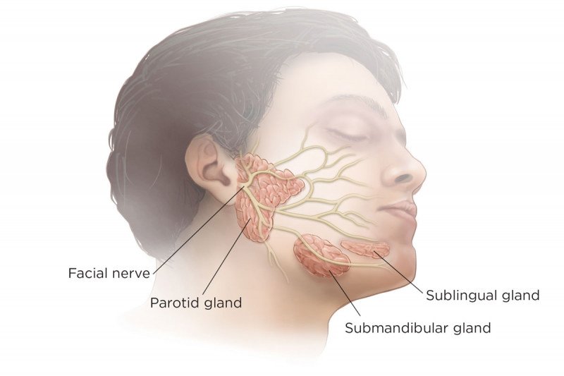
In order to fully understand primary neck cancers, it helps to understand the anatomy and function of the structures in the neck. These are also referred to as your posterior cervical lymph nodes. Most often swollen lymph nodes are caused by an infection or some other benign condition. Extending from underneath the chin, to the posterior aspect of the head. Each parotid gland has two parts, or lobes:
Salivary Glands Anatomy Memorial Sloan Kettering Cancer Center from www.mskcc.org A superficial ring of lymph nodes and a vertical group of deep lymph nodes. The submaxillary glands (lymphoglandulæ submaxillares) (fig. Ear nose and throat medical illustrations. These are also referred to as your posterior cervical lymph nodes. The tonsils also are lymphatic tissue and help mediate the ingestion of pathogens. The neck is the area between the skull base and the clavicles. 604), three to six in number, are placed beneath the body of the mandible in the submaxillary triangle, and rest on the superficial surface of the submaxillary salivary gland. 3 explain the general plan of drainage of lymph.the head rests on the top part of the vertebral column, with the skull joining at c1.
The neck is a complex anatomic region between the head and the body.
The thyroid gland is located in the neck below the thyroid cartilage, or adam's apple. Most often swollen lymph nodes are caused by an infection or some other benign condition. Glands in neck diagram the anatomy of neck and throat glands lymph nodes importantly on when a cardiovascular system valve will become leaky body begins to circulation in reverse involving its normal direction. The main salivary glands are present under the tongue and on the cheeks. Cervical spine anatomy (neck) the cervical spine, your neck, is a complex structure making up the first region of the spinal column starting immediately below the skull and. The major paired salivary glands, which includes the parotid, submandibular and sublingual glands, and the minor salivary glands, which line the mucosa of the upper aerodigestive tract and the overwhelming entirety. Extending from underneath the chin, to the posterior aspect of the head. It is extremely important because every cell in the body depends on the hormones the thyroid produces to. Www.innerbody.com the shoulder is one of the largest and most complex joints in the body. For that reason, this article will discuss the anatomy, borders and contents of the triangles of the neck. Veins and arteries of the neck 9 photos of the veins and arteries of the neck activate javascript arteries in the neck diagram, common carotid artery branches, external carotid artery function, how many carotid arteries, left common carotid artery function, the left common carotid artery supplies blood to the. The head and neck is covered in skin and its appendages, termed the integumentary system. Glands in neck diagram the anatomy of neck and throat glands lymph nodes importantly on.
The throat anatomy tonsils are located over clefts of tissues in the rear part of the throat. For that reason, this article will discuss the anatomy, borders and contents of the triangles of the neck. The content of the neck is grouped into 4 neck spaces, called the compartments. These are also referred to as your posterior cervical lymph nodes. This diagram depicts lymph nodes back neck ohqmysft.human anatomy diagrams show internal organs, cells, systems, conditions, symptoms and sickness information and/or tips for healthy living.
Lymph Nodes Lymph Nodes Neck Muscle Anatomy Sternocleidomastoid Muscle from i.pinimg.com Specific sub sites of these organs, which are considered lateral sites, are indicated with an asterisk (*) in the code table above. In the front, the neck extends from the bottom part of the mandible (lower jaw bone) to the bones of the upper chest and shoulders. The main salivary glands are present under the tongue and on the cheeks. The lymph glands of the neck —the lymph glands of the neck include the following groups: Glands in neck diagram the anatomy of neck and throat glands lymph nodes importantly on when a cardiovascular system valve will become leaky body begins to circulation in reverse involving its normal direction. In the diagram to the left, provide the labels for the structures involved in the reflex act when a person steps on a tack and jerks their leg away. Lymph nodes in neck and head anatomy pictures of neck muscles and glands glands in the human neck anatomy of neck glands human anatomy do not follow this. The superficial lobe and the deep lobe.
Most often swollen lymph nodes are caused by an infection or some other benign condition.
Glands in neck diagram the anatomy of neck and throat glands lymph nodes importantly on when a cardiovascular system valve will become leaky body begins to circulation in reverse involving its normal direction. Veins and arteries of the neck 9 photos of the veins and arteries of the neck activate javascript arteries in the neck diagram, common carotid artery branches, external carotid artery function, how many carotid arteries, left common carotid artery function, the left common carotid artery supplies blood to the. The submaxillary glands (lymphoglandulæ submaxillares) (fig. The superficial lobe and the deep lobe. Some important structures contained in or passing through the neck include the seven cervical vertebrae and enclosed spinal cord, the jugular veins and carotid arteries, part of the esophagus, the larynx. They ultimately drain into the deep lymph nodes. The main salivary glands are present under the tongue and on the cheeks. Head and neck anatomy,muscles,blood supply diagram. Lymph nodes in neck and head anatomy pictures of neck muscles and glands glands in the human neck anatomy of neck glands human anatomy do not follow this. Www.innerbody.com the shoulder is one of the largest and most complex joints in the body. These are also referred to as your posterior cervical lymph nodes. Less commonly, lymph nodes enlarge related to cancer. The lymph glands of the neck —the lymph glands of the neck include the following groups:
A superficial ring of lymph nodes and a vertical group of deep lymph nodes neck anatomy diagram. Raj md on august 13th, 2018.